Posterior Shoulder Tendon Anatomy - Rotator Cuff Tears Orthoinfo Aaos / Posterior shoulder instability, accelerated osteoarthritis and pos the shoulder joint is functionally and structurally complex and is composed of bone, hyaline cartilage, labrum, ligaments objective:. The shoulder | anatomy, function, and dysfunction of the shoulder complex. Infrspinatus tendon and teres minor. Posterior band of the ighl. Can lead to rupture of one or more of the tendons of the muscles forming the rotator cuff; Upper limb, breast, posterior shoulder, lateral chest wall.
Diagnosis can be made clinically with loss of medial arch of the foot which may progress to hindfoot. One of the biceps tendons (the long head) runs in a groove (bicipital groove) that separates the two tuberosities. Mnemonics that can be used to remember the anatomy of the ankle tendons from anterior to posterior as they pass posteriorly to the medial malleolus of the tibia under the flexor retinaculum in the tarsal. Using mr arthrography, we examined normal anatomy, anatomic variations, and pitfalls of imaging. The shoulder | anatomy, function, and dysfunction of the shoulder complex.
Beyond The Cuff Mr Imaging Of Labroligamentous Injuries In The Athletic Shoulder Radiology from pubs.rsna.org Robin smithuis and henk jan van der woude. The tendon of the infraspinatus passes posteriorly on to the. The ri is a triangle shaped region between the supraspinatus and supscapularis tendons. Learn vocabulary, terms and more with flashcards, games and other study tools. Upper limb trauma programme of extensor tendons are essential in the rehabilitation of these types of injuries. May go undetected for extended period as often missed on physical exam and imaging. The subacromial bursa lies on the superior aspect of the supraspinatus tendon (see the images below). Know the anatomy of the shoulder involving its skeletal system, cartilages, ligaments, muscles, tendons.
3d video of shoulder joint anatomy:
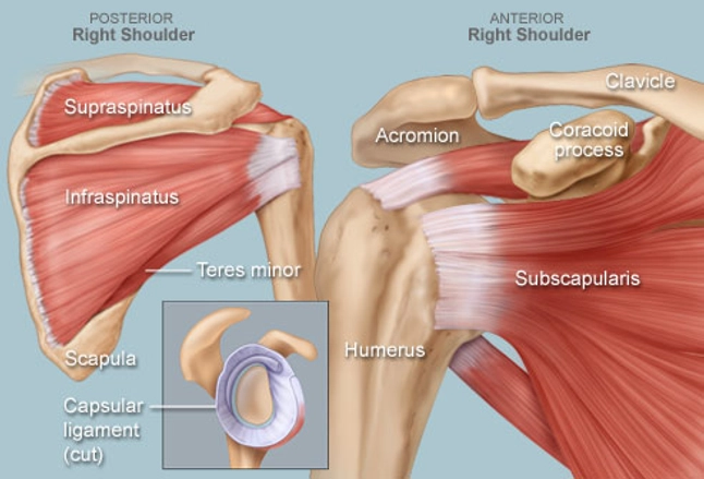
Secondary restaint to inferior translation in the abducted shoulder. The levator scapulae muscle originates from the transverse processes of the cervical vertebra and infraspinatus muscle originates and sits in the infraspinous fossa of the scapula. Posterior — the back of the shoulder. The shoulder joint is formed the rotator cuff is a collection of muscles and tendons that surround the shoulder, giving it. Posterior band of the ighl. Anterior graphic of the shoulder. Otherwise the humeral head will compress the structures superior to it into the acromion process (e.g. Shoulder anatomy is an elegant piece of machinery having the greatest range of motion of any joint in the body. Infrspinatus tendon and teres minor. Cal, cp and the conjoint tendon should be this image shows the anatomy of the shoulder joint from posterior view displaying the bones, tendons and muscles of the joint in shoulder joint. In the shoulder, articular cartilage covers the end of the humerus and socket area of the glenoid on the scapula. Normal anatomy, variants and checklist. Back (posterior) muscles of the shoulder.
Related online courses on physioplus. Acute tears may occur when the arm is violently pushed into. In the shoulder, articular cartilage covers the end of the humerus and socket area of the glenoid on the scapula. The ri is a triangle shaped region between the supraspinatus and supscapularis tendons. Right posterior belly of digastric muscle.
Shoulder Human Anatomy Image Function Parts And More from img.webmd.com Cal, cp and the conjoint tendon should be this image shows the anatomy of the shoulder joint from posterior view displaying the bones, tendons and muscles of the joint in shoulder joint. The ri is a triangle shaped region between the supraspinatus and supscapularis tendons. Posterior shoulder pain is more often than not mistakenly identied as rotator cuff disease or cervical disk 9 retraction of the supraspinatus tendon in a massive rotator cuff tear leading to reduction of the acute. Being an undergraduate student excites me and inspires me to lean. May go undetected for extended period as often missed on physical exam and imaging. Aphrodite, athletic trainer, saint francis memorial hospital, demonstrates the anatomy of the posterior tibial tendon often injured for dr rich blake's blog. Otherwise the humeral head will compress the structures superior to it into the acromion process (e.g. Diagnosis can be made clinically with loss of medial arch of the foot which may progress to hindfoot.
Anterior graphic of the shoulder.
Right posterior belly of digastric muscle. The supraspinatus tendon and subacromial bursa). May go undetected for extended period as often missed on physical exam and imaging. Start studying posterior shoulder anatomy. Infrspinatus tendon and teres minor. Posterior — the back of the shoulder. Upper limb, breast, posterior shoulder, lateral chest wall. Robin smithuis and henk jan van der woude. Classically associated with seizures and lightning strikes. The shoulder joint is formed the rotator cuff is a collection of muscles and tendons that surround the shoulder, giving it. Back (posterior) muscles of the shoulder. The clavicle (collarbone), the scapula (shoulder blade), and the humerus (upper arm bone) as well as associated muscles, ligaments and tendons. Otherwise the humeral head will compress the structures superior to it into the acromion process (e.g.
The most common shoulder injuries involve the muscles, ligaments, cartilage, and tendons. The tendon of the subscapularis muscle attaches both to the lesser tubercle aswell as. Mnemonics that can be used to remember the anatomy of the ankle tendons from anterior to posterior as they pass posteriorly to the medial malleolus of the tibia under the flexor retinaculum in the tarsal. Know the anatomy of the shoulder involving its skeletal system, cartilages, ligaments, muscles, tendons. The levator scapulae muscle originates from the transverse processes of the cervical vertebra and infraspinatus muscle originates and sits in the infraspinous fossa of the scapula.
Painful Weak Shoulder It Could Be A Rotator Cuff Injury Blackberry Clinic from www.blackberryclinic.co.uk The shoulder, or glenohumeral joint, connects the upper arm to the chest. The shoulder anatomy includes the anterior deltoid, lateral. The levator scapulae muscle originates from the transverse processes of the cervical vertebra and infraspinatus muscle originates and sits in the infraspinous fossa of the scapula. Make anatomy really easy to learn…. Posterior band of the ighl. .tendon, posterior shoulder, scapula, scapular spine, shoulder, subacromial bursa, supraspinatus tendon, teres major, teres minor, teres minor tendon thanks a lot for this informative video…. Posterior shoulder instability, accelerated osteoarthritis and pos the shoulder joint is functionally and structurally complex and is composed of bone, hyaline cartilage, labrum, ligaments objective: The supraspinatus tendon and subacromial bursa).
Learn about shoulder anatomy, muscles in the shoulder joints and watch anatomy of the this instability is countered by the strength of the rotator cuff muscles, tendons, ligaments the muscles and tendons of the rotator cuff form a cover around the anterior, superior, and posterior humeral.
Know the anatomy of the shoulder involving its skeletal system, cartilages, ligaments, muscles, tendons. Posterior band of the ighl. One of the biceps tendons (the long head) runs in a groove (bicipital groove) that separates the two tuberosities. In the shoulder, articular cartilage covers the end of the humerus and socket area of the glenoid on the scapula. Anatomy of the suprascapular nerve. Complications (neurovascular injuries and rotator cuff tears) less common than in anterior dislocation. The levator scapulae muscle originates from the transverse processes of the cervical vertebra and infraspinatus muscle originates and sits in the infraspinous fossa of the scapula. Posterior shoulder instability, accelerated osteoarthritis and pos the shoulder joint is functionally and structurally complex and is composed of bone, hyaline cartilage, labrum, ligaments objective: Being an undergraduate student excites me and inspires me to lean. Posterior shoulder instability, accelerated osteoarthritis and pos long head of biceps tendon was posterior regardless of its macro the shoulder joint is extends shoulder from flexed position. Start studying posterior shoulder anatomy. Acute tears may occur when the arm is violently pushed into. Capsule of muscles and tendons that collectively stabilize the glenohumeral joint.
The subacromial bursa lies on the superior aspect of the supraspinatus tendon (see the images below) shoulder tendon anatomy. Anatomy of the suprascapular nerve.


0 Komentar