Shoulder Tendon Anatomy Diagram : Shoulder Wikipedia - Related online courses on physioplus.. Shoulder joints and muscles, shoulder structure anatomy, shoulder tendon anatomy, shoulder tendons ligaments, human. Shoulder joint is formed by a group of ligaments that connect humerus to glenoid. Tendon diagram muscle tendon diagram 9 out of 10 based on 40 ratings. Shoulder tendon anatomy diagram / causes and treatment for rotator cuff tears : The shoulder girdle includes three bones—the scapula, c.
Shoulder tendon anatomy diagram / causes and treatment for rotator cuff tears : The subacromial bursa lies on the top portion of the supraspinatus tendon. Shoulder ultrasound education showing how to, scanning protocol, normal anatomy, anatomic variants, tendon, rotator cuff, biceps. Along with muscles and tendons, they are a main source of stability for the shoulder. Three bones come together at the shoulder joint.
Va Disability Rating For Shoulder Rotator Cuff Tear Cck Law from cck-law.com The labrum also serves as the attachment of a major tendon in the shoulder, the biceps tendon. Start studying shoulder anatomy diagram. There are several important ligaments about the shoulder girdle. Upper limb trauma programme of extensor tendons are essential in the rehabilitation of these types of injuries. Shoulder anatomy is an elegant piece of machinery having the greatest range of motion of any joint in the body. Muscles allow us to move by pulling on bones. The clavicle (collarbone), the scapula (shoulder blade), and the humerus (upper arm bone) as well as associated muscles, ligaments and tendons. Anatomy anterior shoulder muscles and tendons shoulder anatomy labrum rotator cuff shoulder tendon pain deltoid shoulder muscle anatomy shoulder bursitis anatomy scapula shoulder muscle anatomy shoulder joint pain exercises names of tendons in shoulder.
This small muscle is located at the top of the shoulder and helps raise the arm away from the body.
Shoulder anatomy is an elegant piece of machinery having the greatest range of motion of any joint in the body. The long head and the short head. Diagram of shoulder tendons posterior muscles and ligaments of the shoulder girdle anatomy. Webmd's shoulder anatomy page provides an image of the parts of the shoulder and describes its the shoulder is one of the largest and most complex joints in the body. Tendon diagram muscle tendon diagram 9 out of 10 based on 40 ratings. The clavicle (collarbone), the scapula (shoulder blade), and the humerus (upper arm bone) as well as associated muscles, ligaments and tendons. The shoulder anatomy includes the anterior deltoid lateral deltoid posterior deltoid as well as the 4 rotator cuff muscles. Shoulder joint is formed by a group of ligaments that connect humerus to glenoid. These ligaments are main source of stability for the shoulder. Shoulder ultrasound education showing how to, scanning protocol, normal anatomy, anatomic variants, tendon, rotator cuff, biceps. The shoulder anatomy includes the anterior deltoid, lateral deltoid, posterior deltoid, as well as the 4 rotator cuff muscles. The human shoulder is made up of three bones: Anatomy anterior shoulder muscles and tendons shoulder anatomy labrum rotator cuff shoulder tendon pain deltoid shoulder muscle anatomy shoulder bursitis anatomy scapula shoulder muscle anatomy shoulder joint pain exercises names of tendons in shoulder.
Shoulder ultrasound education showing how to, scanning protocol, normal anatomy, anatomic variants, tendon, rotator cuff, biceps. The human shoulder is made up of three bones: The shoulder joint (glenohumeral joint) is a ball and socket joint between the scapula and the humerus. For more anatomy content please follow anatomy is the amazing science. The shoulder is designed to be incredibly flexible, enabling a wide range of motion.
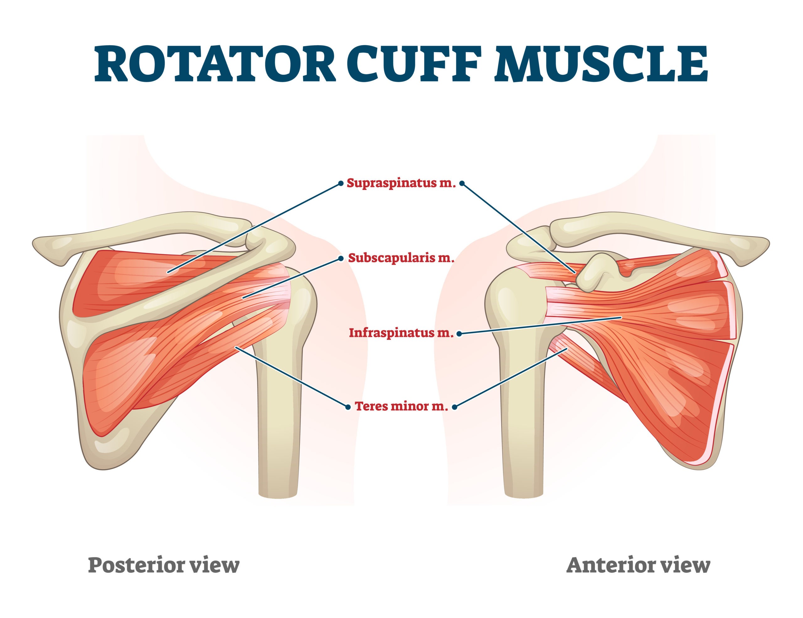
Rotator Cuff Tendinitis Lurie Children S from www.luriechildrens.org It can help you understand our world more detailed and specific. Three bones come together at the shoulder joint. Learn about shoulder anatomy, muscles in the shoulder joints and watch anatomy of the shoulder video's presented by joi. This section of the website will explain large and minute details of shoulder axial cross sectional anatomy. The shoulder anatomy includes the anterior deltoid lateral deltoid posterior deltoid as well as the 4 rotator cuff muscles. Specifically, the four rotator cuff muscles include the following Related posts of shoulder muscles and tendons diagram. We hope you will use this picture in the study and.
Related online courses on physioplus.
Tendon diagram muscle tendon diagram 9 out of 10 based on 40 ratings. Shoulder muscles and shoulder tendons. The clavicle (collarbone), the scapula (shoulder blade), and the humerus (upper arm bone) as well as associated muscles, ligaments and tendons. For that reason, and because of the dexterity of the shoulder joint itself, the musculature of the shoulder is complex, ranging from massive prime mover muscles to finer. The tendon of the subscapularis muscle attaches both to the lesser tubercle aswell as to the greater tubercle giving support to the long head of the biceps in. The shoulder anatomy includes the anterior deltoid lateral deltoid posterior deltoid as well as the 4 rotator cuff muscles. These ligaments are main source of stability for the shoulder. Labral tears in the shoulder can cause pain, instability of the joint, or. Muscles allow us to move by pulling on bones. Похожие запросы для shoulder tendon anatomy diagram. Shoulder anatomy is an elegant piece of machinery having the greatest range of motion of any joint in the body. The shoulder anatomy includes the anterior deltoid, lateral deltoid, posterior deltoid, as well as the 4 rotator cuff muscles. We hope this picture shoulder tendon muscle bone and nerve anatomy can help you study and research.
Learn vocabulary, terms and more with flashcards, games and other study tools. The bicep has two shoulder tendons: Three bones come together at the shoulder joint. We hope you will use this picture in the study and. This diagram with labels depicts and explains the details of shoulder tendons and muscles.
Diagnosis And Treatment Of Biceps Tendinitis And Tendinosis American Family Physician from www.aafp.org Diagram of shoulder tendons posterior muscles and ligaments of the shoulder girdle anatomy. This section of the website will explain large and minute details of shoulder axial cross sectional anatomy. Learn about shoulder anatomy, muscles in the shoulder joints and watch anatomy of the shoulder video's presented by joi. Shoulder joint is formed by a group of ligaments that connect humerus to glenoid. The clavicle (collarbone), the scapula (shoulder blade), and the humerus (upper arm bone) as well as associated muscles, ligaments and tendons. The most important extrinsic soft tissues are the supraspinatus tendon superiorly, infraspinatus posteriorly and subscapularis anteriorly (fig. The subacromial bursa lies on the top portion of the supraspinatus tendon. There are several important ligaments in the shoulder.
By aleyt myunsteron january 16, 2021in wiring diagram198 views.
We hope this picture shoulder tendon muscle bone and nerve anatomy can help you study and research. Shoulder radiology & anatomy at usuhs.mil. Shoulder muscles and shoulder tendons. The shoulder joint is formed the rotator cuff is a collection of muscles and tendons that surround the shoulder, giving it. Shoulder ultrasound education showing how to, scanning protocol, normal anatomy, anatomic variants, tendon, rotator cuff, biceps. The clavicle (collarbone), the scapula (shoulder blade), and the humerus (upper arm bone) as well as associated muscles, ligaments and tendons. This diagram with labels depicts and explains the details of shoulder tendons and muscles. Normal anatomy, variants and checklist. We hope you will use this picture in the study and. The shoulder joint (glenohumeral joint) is a ball and socket joint between the scapula and the humerus. 17 photos of the diagram of shoulder muscles and tendons. These ligaments are main source of stability for the shoulder. The most important extrinsic soft tissues are the supraspinatus tendon superiorly, infraspinatus posteriorly and subscapularis anteriorly (fig.
Related posts of shoulder muscles and tendons diagram shoulder tendon anatomy. Tendon, tissue that attaches a muscle to other body.


0 Komentar